Hemangiomas
Infantile hemangiomas are common benign vascular tumors that develop during early infancy. While many lesions resolve gradually without intervention, early evaluation is essential for hemangiomas in sensitive locations or those at risk of complications.
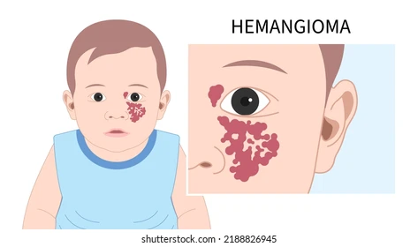
Overview
An infantile hemangioma is a benign vascular tumor that develops during early infancy.
It may not be obvious at birth, or may initially appear as a small mark before beginning to grow during the following weeks.
Infantile hemangiomas generally pass through two broad phases:
- A proliferative phase during which the lesion grows.
- A gradual involution phase over subsequent years.
The most rapid growth commonly occurs between approximately 1 and 3 months of age, and most growth has usually occurred by about 5 months.
Recognizing high-risk hemangiomas early is therefore important because waiting until growth has finished may miss an important treatment window.
In contrast, many small uncomplicated hemangiomas can simply be observed.
A benign vascular tumor developing in early infancy
Fastest growth usually occurs during the first months
Based on location, size, growth and functional risk
Many uncomplicated lesions require observation only
What are Infantile Hemangiomas?
The term "vascular birthmark" is sometimes used broadly for several different vascular conditions, but these conditions are not all hemangiomas.
Infantile hemangiomas differ from vascular malformations, which result from abnormal development of blood vessels.
An infantile hemangioma characteristically becomes visible or begins growing during early infancy, enters a proliferative phase and later gradually involutes.
Hemangiomas may be:
- Superficial, producing a bright red lesion on the skin.
- Deep, creating a skin-colored or bluish swelling beneath the surface.
- Mixed, with both superficial and deeper components.
A child may have a single hemangioma or multiple lesions.
Appearance alone does not determine clinical importance.
Location, growth pattern and potential effect on surrounding structures are major parts of risk assessment.
How do Hemangiomas Develop?
Most infantile hemangiomas first undergo a period of growth and later begin natural involution.
Growth is particularly active during early infancy.
A lesion may therefore change noticeably over a relatively short period during the first few months.
After the proliferative phase slows, the hemangioma generally stabilizes and begins to regress gradually.
Its color may become lighter and its volume may decrease over time.
Although many hemangiomas involute substantially, residual changes can remain, including:
- Excess skin.
- Changes in skin texture.
- Visible superficial blood vessels.
- Altered pigmentation.
- Scarring following previous ulceration.
Treatment decisions therefore consider not only whether a hemangioma may eventually shrink but also whether growth could cause functional impairment or permanent disfigurement before involution occurs.
When is Early Assessment Important?
Not all infantile hemangiomas carry the same level of risk.
Early assessment is particularly important when a hemangioma is:
- Near the eye or interfering with eye opening or vision.
- On the nose, lips, ear or another facial location where growth could cause significant distortion.
- Inside the mouth or near the airway.
- In the beard distribution or lower face when airway involvement may be a concern.
- Large or growing rapidly.
- Ulcerated or repeatedly bleeding.
- Painful.
- Interfering with feeding, breathing, hearing or another important function.
- Large and segmental across a broad area of the face or scalp.
- Large in the lower back, sacral or diaper region.
- Associated with findings that raise concern for underlying congenital abnormalities.
The presence of multiple skin hemangiomas may also prompt further assessment.
When an infant has five or more cutaneous hemangiomas, evaluation of the liver may be recommended to look for internal hemangiomas.
Early evaluation is particularly valuable because the fastest phase of hemangioma growth occurs during early infancy.
Ulceration and Possible Complications
Most infantile hemangiomas do not cause serious complications.
Problems can occur, however, depending on the lesion's location, size and growth pattern.
Ulceration is an important complication.
It occurs when the skin covering the hemangioma breaks down and forms an open sore.
Ulceration may cause:
- Pain.
- Bleeding.
- Difficulty with wound care.
- Risk of infection.
- Residual scarring.
Hemangiomas affecting important organs or functions require particular attention.
A lesion near the eye may interfere with visual development if it blocks vision or distorts the eye.
Hemangiomas around the mouth may interfere with feeding.
Airway hemangiomas or lesions affecting the airway can cause breathing problems and require prompt specialist assessment.
Large or specifically distributed hemangiomas in certain body regions may sometimes be associated with underlying structural abnormalities requiring additional investigation.
Diagnosis
Most infantile hemangiomas are diagnosed from the clinical history, appearance of the lesion and physical examination.
The clinician may ask:
- Was the mark visible at birth?
- When did growth begin?
- How rapidly has it changed?
- Has there been ulceration, bleeding or pain?
- Does it affect vision, feeding, breathing or another function?
- Are there other hemangiomas on the skin?
Imaging is not required routinely for every hemangioma.
Imaging may be considered when:
- The diagnosis is uncertain.
- Five or more cutaneous hemangiomas are present.
- Associated internal or structural abnormalities are suspected.
Ultrasound can be used as an initial imaging study when the diagnosis is uncertain.
MRI may be required when a large or specifically distributed hemangioma raises concern for associated structural abnormalities.
Investigations should therefore be directed toward a specific clinical question rather than performed routinely for every vascular lesion.
Treatment
Many infantile hemangiomas do not require active treatment.
A small lesion that is not ulcerated, does not threaten an important function and is unlikely to cause significant permanent disfigurement may be managed with observation and follow-up.
When a hemangioma is high risk or causing a problem, treatment options depend on the individual lesion.
Propranolol:
Oral propranolol is the first-line systemic medication for infantile hemangiomas that require systemic treatment.
It can slow growth and promote regression.
Because propranolol affects the whole body, it must be prescribed and monitored by an appropriate medical team after the child has been assessed.
Assessment may include review of:
- Medical history.
- Cardiovascular health.
- Respiratory health.
- Feeding.
- Other medications.
Parents should follow the prescribed plan carefully and should not change or stop treatment without medical guidance.
Timolol:
Topical timolol may be considered for selected small, thin and superficial hemangiomas.
It is not appropriate for every lesion.
Laser Treatment:
Laser therapy can be useful in selected circumstances, particularly for certain superficial residual vascular changes or as part of individualized treatment.
Surgery:
Most infantile hemangiomas do not require surgical removal during the proliferative phase.
Surgery may be appropriate in selected cases and may also be used later to correct excess skin or residual deformity after involution.
Treatment selection depends on the lesion's location, stage, associated risks and effect on the child.
Propranolol Treatment and Follow-up
When propranolol is selected, treatment should be supervised medically.
Before treatment begins, the clinician assesses whether the medication is appropriate for the child, paying particular attention to the medical history and cardiovascular and respiratory examination.
Possible adverse effects can include:
- Reduced heart rate.
- Reduced blood pressure.
- Low blood glucose.
- Sleep disturbance.
- Respiratory symptoms in some children.
Families receive specific instructions about administering propranolol in relation to feeding.
During illnesses associated with reduced oral intake or vomiting, the medical team may instruct parents to temporarily withhold doses to reduce the risk of hypoglycemia.
Families should follow the individualized instructions provided for their child rather than independently changing treatment.
Follow-up may monitor:
- Hemangioma growth.
- Changes in size and color.
- Treatment response.
- Adverse effects.
- The child's weight because treatment plans may need adjustment as the infant grows.
Treatment duration varies according to the hemangioma, the child's age and response.
When is Surgery Used?
Surgery is not first-line treatment for most infantile hemangiomas.
It may be considered in selected situations when:
- A problem cannot be managed adequately with medication or other options.
- The lesion is in a location where removal can provide an appropriate functional or cosmetic result.
- A particular complication requires intervention.
- Excess skin or deformity remains after involution.
In many cases, surgery or laser treatment is especially useful after substantial involution has occurred to address residual skin or tissue changes.
Timing is individualized.
The decision considers:
- The child's age.
- Hemangioma location.
- Size and depth.
- Whether the lesion is still growing or involuting.
- Potential scarring.
- Functional impact.
- The expected result of observation compared with intervention.
No single surgical rule applies to every hemangioma.
Before a Surgical Procedure
If surgery or another procedure is selected, the medical team assesses both the child and the hemangioma and defines the goal of treatment.
Assessment may include:
- Location and depth of the lesion.
- Stage of growth or involution.
- Response to previous treatment.
- Previous ulceration or scarring.
- Surrounding tissues.
- Functional problems.
- The child's overall health.
Parents should follow fasting and medication instructions when anesthesia is planned.
The medical team should be informed if the child:
- Takes regular medication.
- Is currently using propranolol or another hemangioma treatment.
- Has medication allergies.
- Has a bleeding problem.
- Develops fever, infection or a new illness before the procedure.
- Has cardiac, respiratory or another important medical condition.
Before treatment, parents can discuss:
- The goal of the procedure.
- Alternatives.
- Expected results.
- Potential scarring.
- Wound care.
- Whether further treatment or follow-up may be needed.
After the Procedure and Follow-up
Care after surgery or laser treatment depends on the procedure and the location of the hemangioma.
Parents receive instructions regarding:
- Wound care.
- Medication.
- Bathing.
- Protection from friction or trauma.
- Activity.
- Follow-up.
Some swelling, redness or discomfort can occur during early healing depending on the procedure.
The medical team should be contacted if the child develops:
- Persistent bleeding.
- Severe or increasing pain.
- Increasing redness or swelling.
- Wound drainage or signs of infection.
- Significant fever.
- Separation of the wound.
- Any sudden change that concerns the family.
Follow-up extends beyond initial wound healing when needed.
The child may continue to be assessed for:
- Tissue growth.
- Scar development.
- Residual superficial vessels.
- Function of the affected area.
- Need for additional treatment in the future.
Frequently Asked Questions
Does every hemangioma need treatment?
No. Many infantile hemangiomas are uncomplicated, do not affect function or vital structures, and can be managed with active observation and regular clinical follow-up until they naturally regress.
Is an infantile hemangioma always present at birth?
An infantile hemangioma may not be obvious at birth. A subtle precursor mark may be present, but noticeable growth typically begins during the first few weeks of life.
When do hemangiomas grow fastest?
The most rapid growth phase usually occurs between 1 and 3 months of age, and much of the proliferation is completed by around 5 months in many infants. Early evaluation is therefore important for high-risk lesions.
Is every red vascular birthmark a hemangioma?
No. There are several types of vascular birthmarks and anomalies. Infantile hemangiomas differ from vascular malformations, and correct clinical evaluation is needed to ensure accurate management.
When can a hemangioma be dangerous?
Hemangiomas become more concerning when they impair vision, breathing, feeding or other functions, when they are large, rapidly growing or ulcerated, or when their location or pattern suggests associated anomalies or complications.
Does my child need imaging?
Not usually. Most infantile hemangiomas are diagnosed by clinical history and physical examination. Imaging is reserved for uncertain diagnosis, multiple cutaneous lesions, deep lesions, or when an underlying syndromic association is suspected.
What medication is commonly used when systemic treatment is needed?
Oral propranolol is the standard first-line medication when systemic therapy is indicated. It must be initiated and monitored by a doctor because it can affect heart rate, blood pressure, blood sugar and breathing in certain patients.
Will a hemangioma disappear without leaving any mark?
While many regress substantially, some lesions leave residual skin changes such as fibrofatty tissue, discoloration, telangiectasias or scarring, especially if they were large, thick or ulcerated.
Book Your Consultation
If your child has an infantile hemangioma that requires evaluation or you would like to discuss treatment options, you can schedule a consultation for a detailed clinical assessment and a tailored management plan.
Video Presentation
Video presentation coming soon